East Meets West: Re-imagining Acupoints Through the Lens of Modern Neuroscience
/East Meets West: Re-imagining Acupoints Through the Lens of Modern Neuroscience
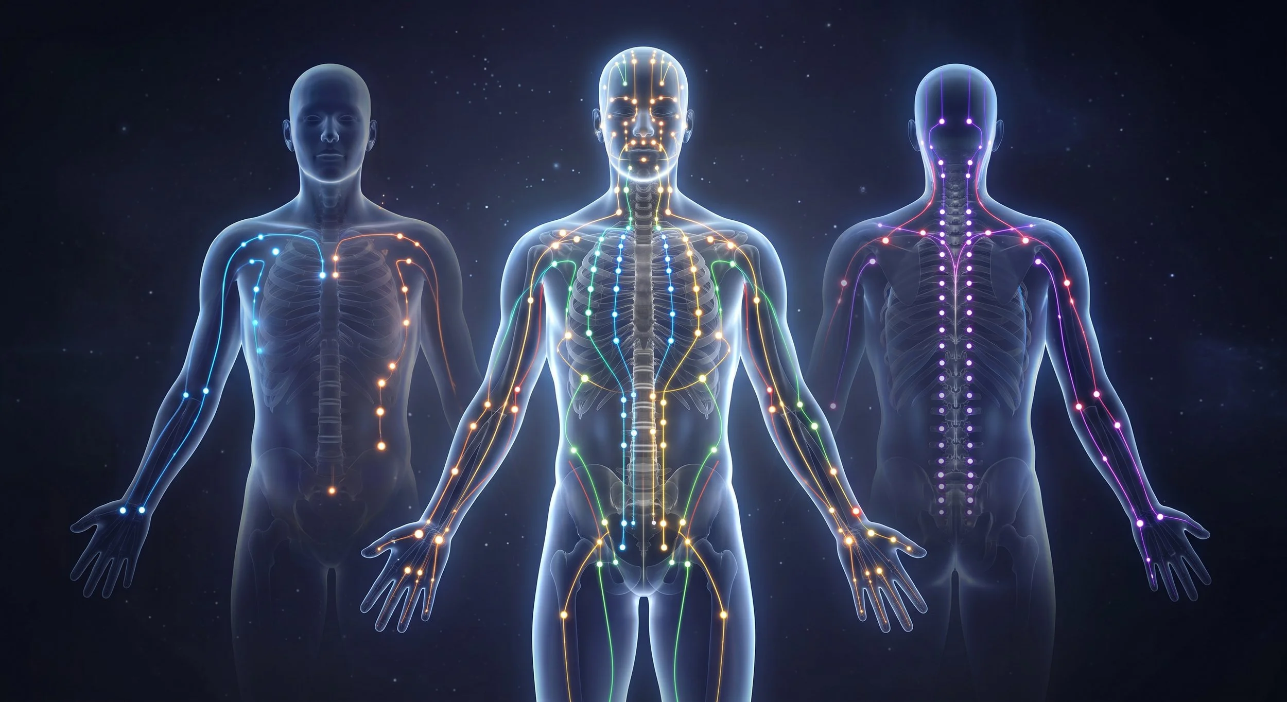
In Traditional Chinese Medicine (TCM), meridians (Jing Luo) are understood as an interconnected network through which energy (Qi), blood, and fluids circulate to sustain bodily function and maintain dynamic homeostatic balance. Rather than originating purely as abstract concepts, early channel maps were rooted in tangible neuroanatomical observations
Moving beyond pre-scientific narratives, medical acupuncture grounds traditional practice in modern neuroanatomy and systems biology. Rather than relying on abstract concepts of metaphysical energy, an integrated framework explains acupuncture through nervous system circuits and tissue dynamics—a shift that opens up promising new avenues for treating chronic pain and chronic non-communicable disease.
While many prominent scientists study the neuroscience of acupuncture, four influential researchers—Huang Longxiang, Qiufu Ma, Alejandro Elorriaga Claraco, and Poney Chiang—stand out for how their work systematically addresses distinct layers of acupuncture mechanics. Rather than discarding traditional concepts, their combined work demonstrates that ancient acupuncture maps were functional diagrams of the human nervous system.
1. Historical & Anatomical Demystification (Huang Longxiang - China Academy of Chinese Medical Sciences, Beijing, China)
Huang Longxiang lays the foundation by re-evaluating classical texts through a historical lens. He argues that early Chinese acupuncturists were observing real anatomical pathways—specifically blood vessels, nerves, and fascial planes—rather than invisible, mystical conduits. His work reconciles ancient meridian terminology with early human dissection, establishing that traditional acupoints were mapped to sensitive neurovascular structures.
Huang, L. X. (2023). [On acupoints]. Zhongguo zhen jiu = Chinese acupuncture & moxibustion, 43(10), 1189–1201. https://doi.org/10.13703/j.0255-2930.20230227-0002
Huang, L. X. (2023). [Evolution of acupuncture techniques for jin diseases and the rise and fall of jingjin doctrine]. Zhongguo zhen jiu = Chinese acupuncture & moxibustion, 43(8), 855–867. https://doi.org/10.13703/j.0255-2930.20230316-k0002
Huang, L. X. (2024). Exploring and detecting the gateway to human body regulation: a perspective from modern anatomy and classical acupuncture. Modern Chinese Clinical Medicine, 31(1): 1-15. https://doi.org/10.3969/j.issn.2095-6606.2024.01.001
Huang, L. X. (2026). [Discovery and rediscovery of morphological structure of human body explored by manual detection and acupuncture practice]. Zhongguo zhen jiu = Chinese acupuncture & moxibustion, 46(8), 1219–1233. https://doi.org/10.13703/j.0255-2930.20250331-0004
Huang, L. X. (2026). [Concept elaboration of shuxue and approval of terminology: interaction with scientific exploration of acupuncture and moxibustion]. Zhongguo zhen jiu = Chinese acupuncture & moxibustion, 46(8), 1303–1318. https://doi.org/10.13703/j.0255-2930.20250617-k0001
2. Cellular & Neuro-autonomic Pathways (Qiufu Ma, Westlake University, Hangzhou, Zhejiang, China)
Qiufu Ma bridges the gap between mechanical needling and systemic regulation. Through neurobiological research, Ma demonstrated that electroacupuncture triggers specific somatosensory-autonomic reflexes depending on location and intensity. For instance, driving low-intensity stimulation at specific nerve-dense regions (like the ST36 acupoint) selectively engages a vagal-adrenal anti-inflammatory pathway, proving that target specificity has a neuroanatomical and somatotopic basis.
Ma, Q. (2020). Somato-Autonomic Reflexes of Acupuncture. Medical acupuncture, 32(6), 362–366. https://doi.org/10.1089/acu.2020.1488
Ma, Q. (2022). A functional subdivision within the somatosensory system and its implications for pain research. Neuron, 110(5), 749–769. https://doi.org/10.1016/j.neuron.2021.12.015
Niruthisard, S., Ma, Q., & Napadow, V. (2024). Recent advances in acupuncture for pain relief. Pain reports, 9(5), e1188. https://doi.org/10.1097/PR9.0000000000001188
Dong, S., Zhao, L., Liu, J., Sha, X., Wu, Y., Liu, W., Sun, J., Su, Y., Zhuang, Z., Chen, J., Dong, Y., Xie, B., Zhou, A., Ji, H., Wang, Y., Deng, X., Jing, X., Ma, Q., Wang, N., & Liu, S. (2025). Neuroanatomical organization of electroacupuncture in modulating gastric function in mice and humans. Neuron, 113(19), 3243–3259.e11. https://doi.org/10.1016/j.neuron.2025.06.023
3. Neurofunctional Model (Alejandro Elorriaga Claraco - McMaster University, Hamilton, Ontario, Canada)
Elorriaga Claraco takes these physiological principles into clinical medicine via the Contemporary Neurofunctional Acupuncture model. Rather than viewing acupoints in isolation, he maps treatment to functional movement systems, target nerve trunks, and segmentally linked organ systems. In his framework, needling acts as a input signal to reset dysfunctional peripheral and central neural feedback loops, treating movement disorders and pain by restoring normal neuromodulation.
4. Precise Peripheral Target Mapping (Poney Chiang- York University, Toronto, Ontario, Canada)
Poney Chiang synthesizes classical point location with modern anatomy. Through neuro-anatomical research, Chiang demonstrates that classical points precisely overlay key nerve structures—such as motor points, cutaneous nerve emergence sites, and nerve trunk crossings through fascial foramina. His Neuro-Meridian Integrative model provides explicit needle techniques to mechanically engage these nerve-fascia junctions, causing local tissue releases and immediate neuro-modulatory responses.
Dorsher, P. T., & Chiang, P. (2018). Neuroembryology of the Acupuncture Principal Meridians: Part 3. The Head and Neck. Medical acupuncture, 30(2), 80–88. https://doi.org/10.1089/acu.2018.1271
Lee, M., Longenecker, R., Lo, S., & Chiang, P. (2019). Distinct Neuroanatomical Structures of Acupoints Kidney 1 to Kidney 8: A Cadaveric Study. Medical acupuncture, 31(1), 19–28. https://doi.org/10.1089/acu.2018.1325
Meltz, L., Ortiz, D., & Chiang, P. (2020). The Anatomical Relationship Between Acupoints of the Face and the Trigeminal Nerve. Medical acupuncture, 32(4), 181–193. https://doi.org/10.1089/acu.2020.1413
Chiang, P., & Martins, M. R. (2022). The Anatomical Specificity Between Acupoints and the Facial Nerve: A Cadaveric Study. Medical acupuncture, 34(6), 391–399. https://doi.org/10.1089/acu.2022.0030
Chiang, P., Chang, D., Moon, H., & Chae, Y. (2023). Neuroanatomical characteristics of the traditional acupuncture point location ST36. Acupuncture in medicine : journal of the British Medical Acupuncture Society, 41(6), 376–377. https://doi.org/10.1177/09645284231200043
Liu, J., & Chiang, P. (2025). Neuroanatomical Specificity of Acupuncture Points of the Shoulder Girdle. Medical acupuncture, 37(5), 398–407. https://doi.org/10.1089/acu.2024.0078
Schreiber, A. D., Chae, Y., & Chiang, P. (2026). Distinct Neuroanatomical Targets of Acupoints Stomach 36—Stomach 41. Medical Acupuncture. https://doi.org/10.1177/19336586261434834
How Their Ideas Form a Unified Framework
Together, these four thinkers form a cohesive, end-to-end medical account of acupuncture:
The Historical Map (Huang Longxiang): Shows that traditional meridians were early anatomical observations of nerve pathways and blood vessels.
The Structural Targets (Poney Chiang): Pinpoints the exact neuro-fascial tissue interfaces where needles interact with sensory nerve endings.
The Biological Signals (Qiufu Ma): Maps how physical stimulation at these specific peripheral sites travels up the spinal cord to trigger distant autonomic and anti-inflammatory reflexes.
The Clinical Application (Alejandro Elorriaga Claraco): Transforms these pathways into a system for treating neuromuscular dysfunction, chronic pain, and systemic imbalance.

Acupoints are often located along the fascial tissues enriched with nerves, vascular/lymphatic vessels, and immune cells. The insertion of an acupuncture needle provides mechanical stimulation of specialized sensory receptors located in the cutaneous and subcutaneous structures. Preferential sites for acupuncture stimulation are associated with areas rich in specialized sensory receptors such as muscle spindles, Golgi tendon organs, ligament receptors, Paciniform and Ruffini’s receptors (joint capsules), deep pressure endings (within muscle belly), and free nerve endings (muscle and fascia). Based on the neurological model, all these areas are highly innervated and as a result there are a number of physiological responses that help modulate the experience of pain. An observed favorable outcome may be explained by overlapping mechanisms in the periphery, spinal cord, and brain.